Description
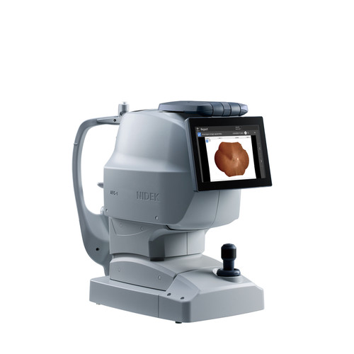
The RetiCam 3100 Plus is an ultra-wide fundus camera built for fast, repeatable retinal documentation across the posterior pole and the far periphery. A single capture records 176° of the retina, and the montage function extends coverage to approximately 220°, bringing the macula, optic disc, vascular arcades, and peripheral retina into one imaging session. True-colour LED illumination reproduces the retina as it appears clinically, avoiding the colour shifts introduced by pseudo-colour imaging methods.
As a non-mydriatic ultra-widefield fundus camera, the RetiCam 3100 Plus images through pupils as small as 3 mm, so most patients can be screened without dilation drops and the added chair time they require. Fully automated alignment, focus, working distance, and exposure support one-button binocular capture, which shortens staff training and keeps imaging throughput consistent in busy clinics.
Onboard software provides gamma, contrast, brightness, and colour adjustment alongside lesion area and cup-to-disc ratio measurement tools, so images captured on the RetiCam 3100 Plus can be reviewed, annotated, and compared across visits.
FEATURES
- Ultra-Widefield Imaging: Capture up to 176° imaging field in a single shot.
- 220° Montage Capture: Montage capability on the RetiCam 3100 Plus ultra-widefield fundus camera extends coverage up to 220°* for comprehensive retinal visualization. 220° field of view is achieved through montage stitching and may vary by clinical and operational factors.
- Non-Mydriatic Operation: Generally without routine dilation, enhancing patient comfort and streamlining the imaging process.
- Automatic Operation: Automated alignment, focus, and exposure for quick image capture without specialized operator training and reduced user error risk.
- True-Colour Illumination: Advanced LED technology delivers accurate colouration, reducing misdiagnosis risks and improving workflow efficiency.
- Flexible Software Tools: Adjust gamma, contrast, brightness, and colour. Easily measure lesion areas and cup-disc ratios.
- Seamless Connectivity: Integrates with DICOM 3.0 for PACS connection. LAN, USB, and FTP support to ensure real-time data sharing and secure backup.
- AI-Powered Positioning: An AI-powered intelligence system enables highly precise positioning for quick fundus image acquisition.
- Minimal Lid and Lash Obstruction: The RetiCam 3100 Plus produces little to no lid or lash obstruction and maintains uniform clarity across all image fields, including the superior and inferior retina.
Frequently Asked Questions
Does the RetiCam 3100 Plus require pupil dilation?
The camera uses non-mydriatic imaging technology, meaning retinal images can generally be captured without pharmacological pupil dilation. Clinical requirements may vary depending on the patient and examination.
How quickly does the camera capture an image?
The RetiCam 3100 Plus has an image capture speed of less than 60 milliseconds per image, helping reduce motion-related challenges during image acquisition.
What file formats does the system produce?
The RetiCam 3100 Plus exports images in standard image file formats, making it easier to incorporate captured images into compatible clinical documentation and data-management workflows.
Is specialized operator training required?
The automated alignment, focus, and exposure functions are designed to simplify image capture and minimize the need for specialized operator training. Appropriate product and clinical training should still be completed before use.